Home

parc jurassique sexcuser Spécialité dapi laser avantage évaporation Réconcilier

Fluorescence microscopy and confocal laser scanning microscopy images... |  Download Scientific Diagram
Fluorescence microscopy and confocal laser scanning microscopy images... | Download Scientific Diagram

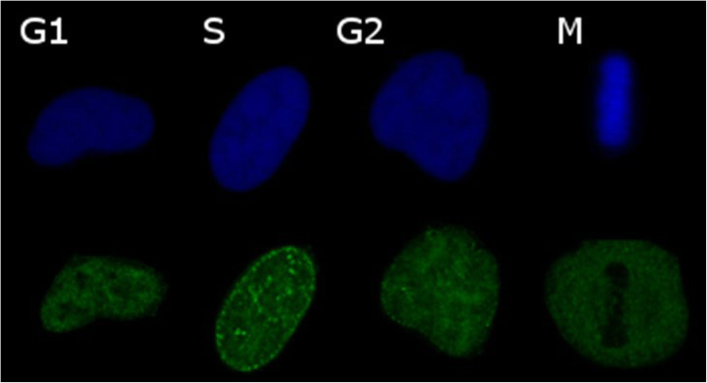
Why Cell Cycle Analysis Details Are Critical In Flow Cytometry - Cheeky  Scientist
Why Cell Cycle Analysis Details Are Critical In Flow Cytometry - Cheeky Scientist

Confocal tutorial: Applications: cross-talk correction2. AMU Dept Pathol  Univ Hel
Confocal tutorial: Applications: cross-talk correction2. AMU Dept Pathol Univ Hel

FadeStop™ Fluorescent Mounting Medium with DAPI #272 | GenuIN Biotech

DAPI - Blue Fluorescent DNA Stain | AAT Bioquest

The use of DAPI fluorescence lifetime imaging for investigating chromatin  condensation in human chromosomes | Scientific Reports
The use of DAPI fluorescence lifetime imaging for investigating chromatin condensation in human chromosomes | Scientific Reports
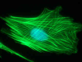
Solved 3. [2 pts] You will be conducting a multi-color | Chegg.com

Cell Viability
Cell Viability

Non-destructive, label free identification of cell cycle phase in cancer  cells by multispectral microscopy of autofluorescence | BMC Cancer | Full  Text
Non-destructive, label free identification of cell cycle phase in cancer cells by multispectral microscopy of autofluorescence | BMC Cancer | Full Text
A) Laser scanning confocal images of the nuclei (DAPI), membrane... |  Download Scientific Diagram
A) Laser scanning confocal images of the nuclei (DAPI), membrane... | Download Scientific Diagram

DAPI (CAS 28718-90-3): R&D Systems

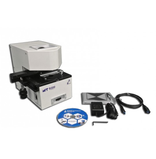
MT-B200/DAPI/Hoechst/Alexa/Fluor350 – Digital Brightfield/Fluorescent  Microscope Imaging System with Integrated Digital Camera
MT-B200/DAPI/Hoechst/Alexa/Fluor350 – Digital Brightfield/Fluorescent Microscope Imaging System with Integrated Digital Camera
Nuclei Watershed Separation - ImageJ

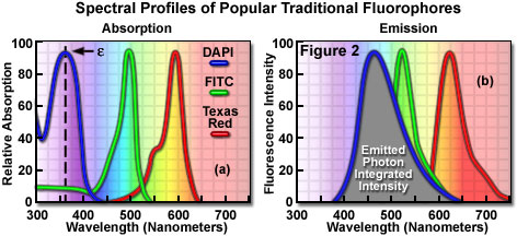
Confocal Microscopy - Fluorophores for Confocal Microscopy | Olympus LS

FadeStop™ Fluorescent Mounting Medium with DAPI #272 | GenuIN Biotech

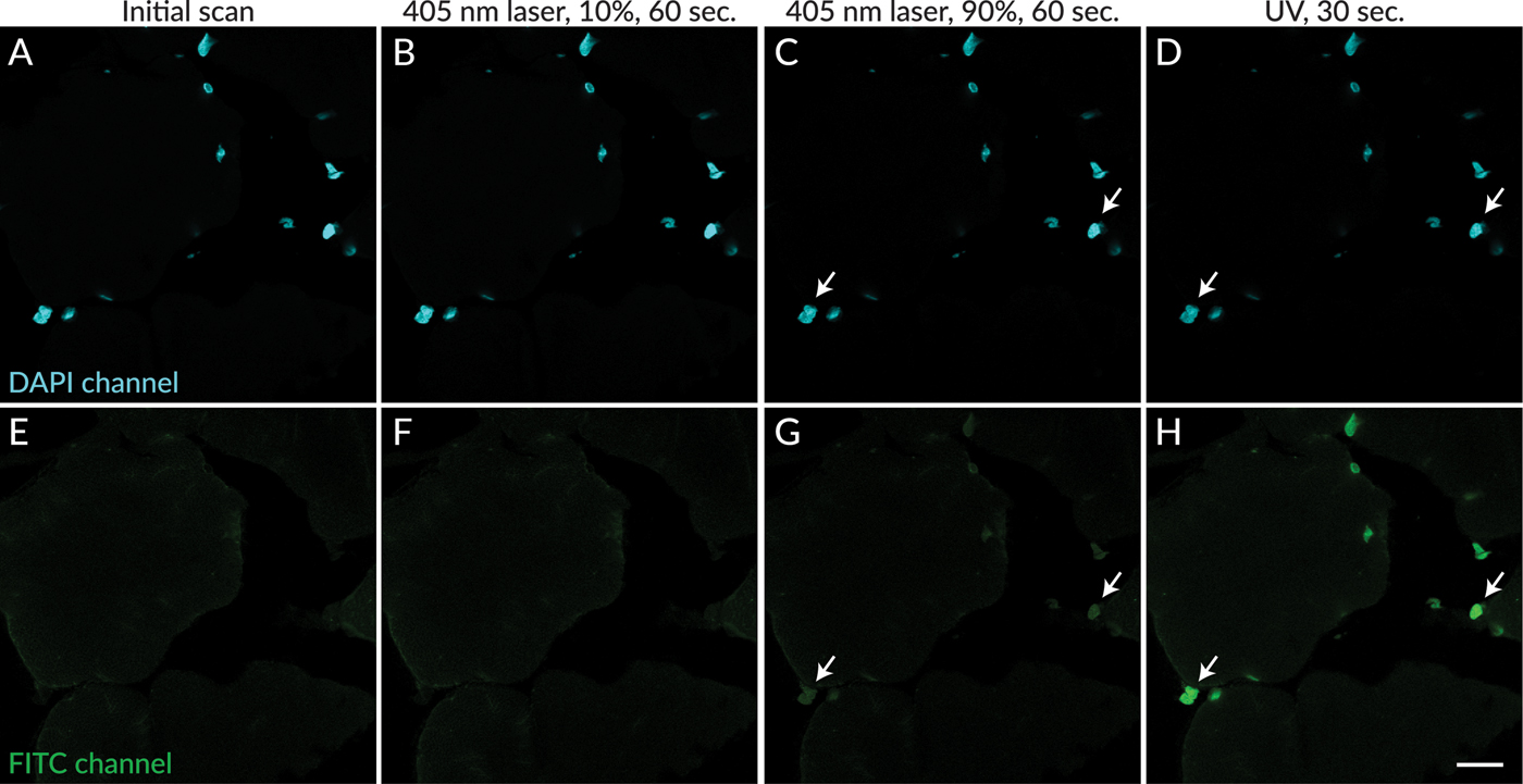
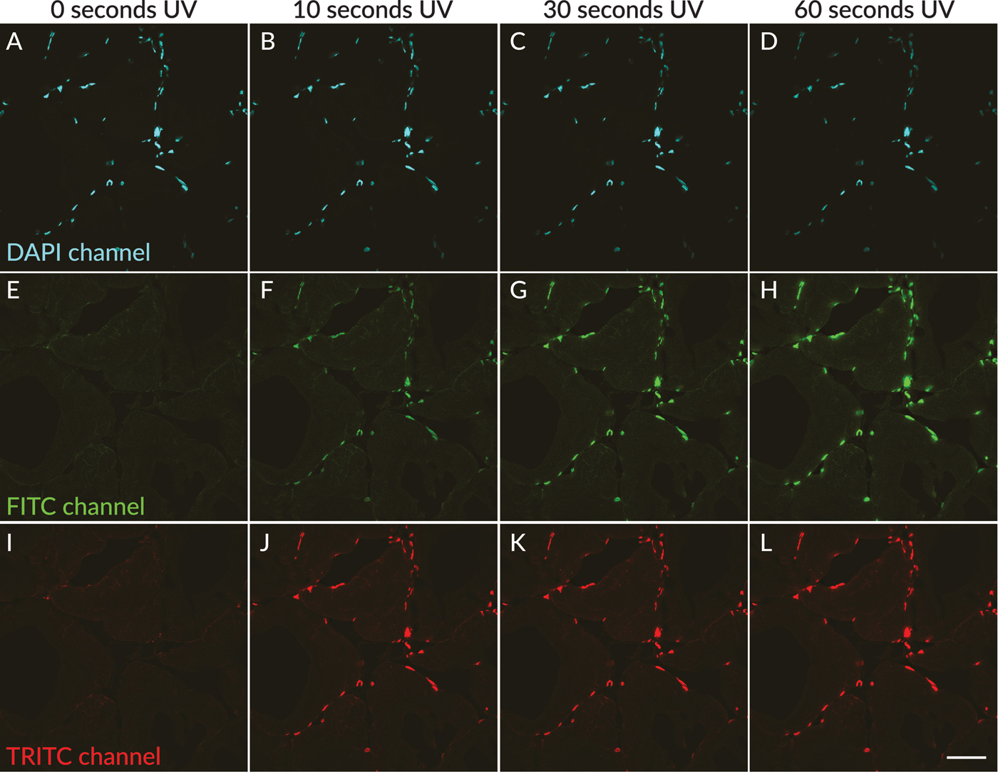
Tech Tip: Avoiding Artifacts from UV Photoconversion of DAPI and Hoechst -  Biotium
Tech Tip: Avoiding Artifacts from UV Photoconversion of DAPI and Hoechst - Biotium

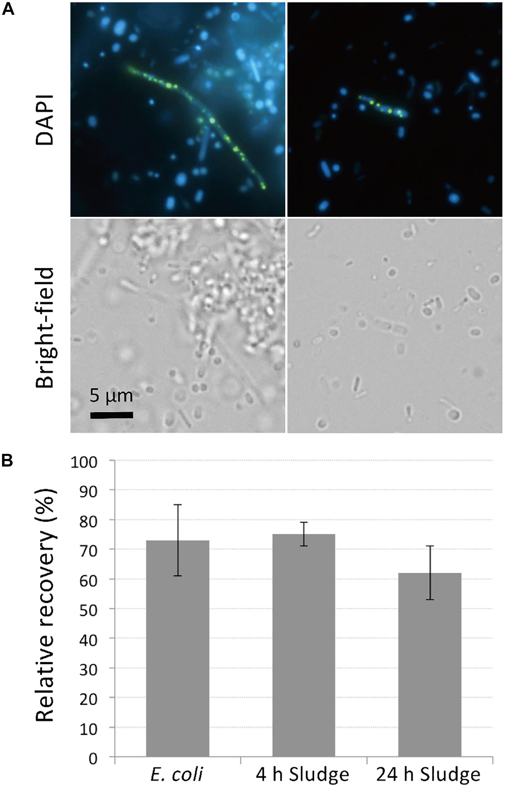
Frontiers | Rapid Enrichment and Isolation of Polyphosphate-Accumulating  Organisms Through 4'6-Diamidino-2-Phenylindole (DAPI) Staining With  Fluorescence-Activated Cell Sorting (FACS)
Frontiers | Rapid Enrichment and Isolation of Polyphosphate-Accumulating Organisms Through 4'6-Diamidino-2-Phenylindole (DAPI) Staining With Fluorescence-Activated Cell Sorting (FACS)

Tips for Avoiding Artifacts from UV Photoconversion of DAPI | Microscopy  Today | Cambridge Core
Tips for Avoiding Artifacts from UV Photoconversion of DAPI | Microscopy Today | Cambridge Core

The excitation and emission spectra of DAPI (4 ʼ... | Download Scientific  Diagram
The excitation and emission spectra of DAPI (4 ʼ... | Download Scientific Diagram

a)). z-Projection of the dapi channel of a complete stack of confocal... |  Download Scientific Diagram
a)). z-Projection of the dapi channel of a complete stack of confocal... | Download Scientific Diagram

Tips for Avoiding Artifacts from UV Photoconversion of DAPI | Microscopy  Today | Cambridge Core
Tips for Avoiding Artifacts from UV Photoconversion of DAPI | Microscopy Today | Cambridge Core

DAPI Solution
DAPI Solution

PhenoVue DAPI nuclear stain | PerkinElmer

DAPI and Confocal Laser-Scanning Microscopy for In Vivo Imaging of  Phytoplasmas | SpringerLink
DAPI and Confocal Laser-Scanning Microscopy for In Vivo Imaging of Phytoplasmas | SpringerLink

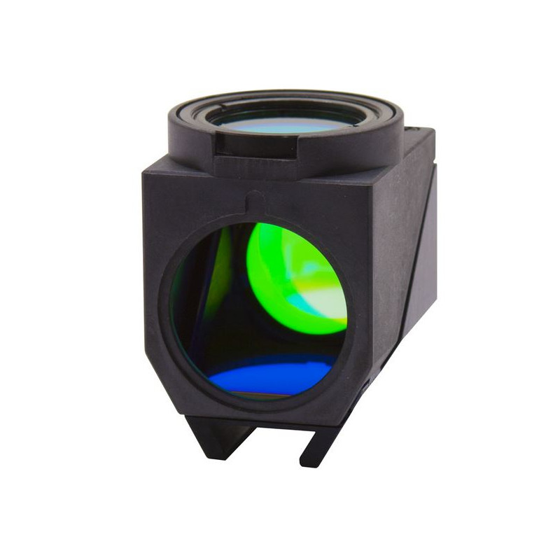
Optika M-1166 fluorescence microscopy filter set, UV-DAPI with filter block  (B-1000 FL HBO)
Optika M-1166 fluorescence microscopy filter set, UV-DAPI with filter block (B-1000 FL HBO)

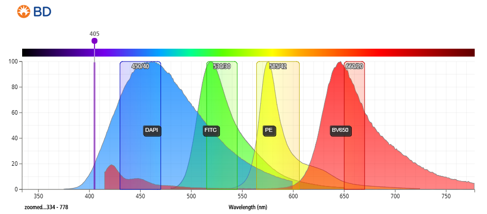
ImageStream®X Mark II Flow Cytometer - Laser Options | Life Science  Research | MilliporeSigma
ImageStream®X Mark II Flow Cytometer - Laser Options | Life Science Research | MilliporeSigma

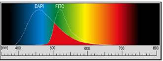
Dual Band Excitation: DAPI-FITC | Nikon's MicroscopyU